A 63-year-old woman’s sudden loss of vision in her right eye led doctors to a Klebsiella brain abscess and a stranger finding: bacteria linked to a prior urinary infection appear to have evolved inside her body into a more invasive form. The case was reported in the New England Journal of Medicine.
According to her doctors, brain MRI scans showed inflammation in the woman’s right eye and lesions in her parietal lobe, a brain region involved in processing sensory information. Blood and urine tests pointed to infection and kidney problems. When surgeons performed a brain biopsy, they found pus, consistent with an abscess.
Cultures from two urine samples and the brain sample all grew Klebsiella pneumoniae, a bacterium that can live in the gastrointestinal tract and cause infections including pneumonia and urinary tract infections. Some forms can spread through the bloodstream to other sites, including the liver, lungs, eyes and brain.
How did UTI bacteria reach her brain?
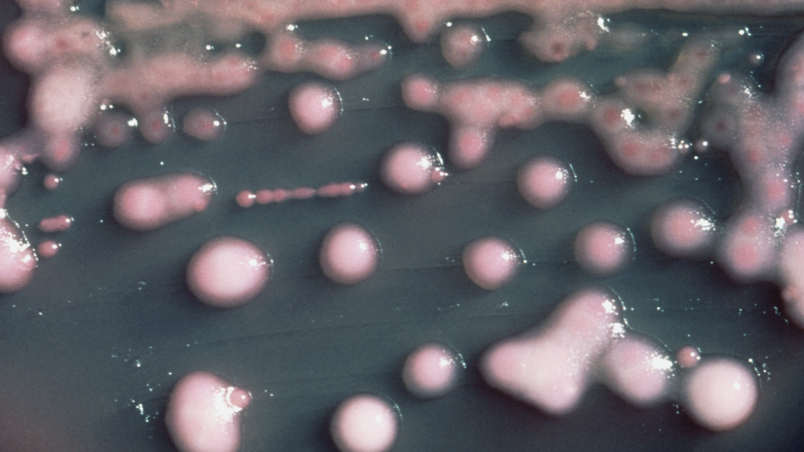
The researchers found that the bacteria were related, but not identical. The first urine sample looked like ordinary K. pneumoniae. The second urine sample and the brain sample produced unusually sticky bacteria with thick capsules, a protective coating around the cell.
That capsule changed the bug’s behavior. In lab testing reported by the doctors and researchers, immune cells called macrophages could not engulf the sticky bacteria as effectively as they could the normal-looking isolate. In mice, the sticky isolates were lethal, while the normal isolate was not.
The tradeoff was ugly but informative. The hypervirulent bacteria grew more slowly and were worse at infecting bladder cells. That suggests they were less fit in the urinary tract, where the infection began, but better equipped to survive in the bloodstream and seed distant organs.
Genome sequencing tied the samples together. The two urine isolates differed by 66 small mutations, while the two hypervirulent isolates differed by just four. The sticky isolates carried mutations in genes involved in building the bacterial capsule. When researchers engineered those mutations into normal K. pneumoniae, the bacteria developed the same sticky phenotype.
The team estimated that the bacteria from the urine samples split from a common ancestor one to two years before the woman’s brain abscess. Her doctors noted that she had been hospitalized with a urinary tract infection two years earlier.
What is heterovirulence?
The researchers described the case as an example of heterovirulence: a mixed bacterial population in one patient where a subpopulation becomes more dangerous without taking over the original site of infection. That differs from heteroresistance, where a resistant subpopulation survives antibiotics but may carry a cost such as slower growth.
There was one useful break in the case. All three bacterial isolates remained susceptible to antibiotics, and doctors were able to treat the infection. The report did not include follow-up details on whether the woman’s neurologic symptoms or vision recovered.
The case shows how K. pneumoniae can persist in a urinary reservoir, evolve new traits and spread to distant body sites, according to the doctors and researchers. It is a reminder that bacterial evolution is not an abstract lab diagram. Sometimes it is happening inside the patient on the table.
This story draws on original reporting from Ars Technica.