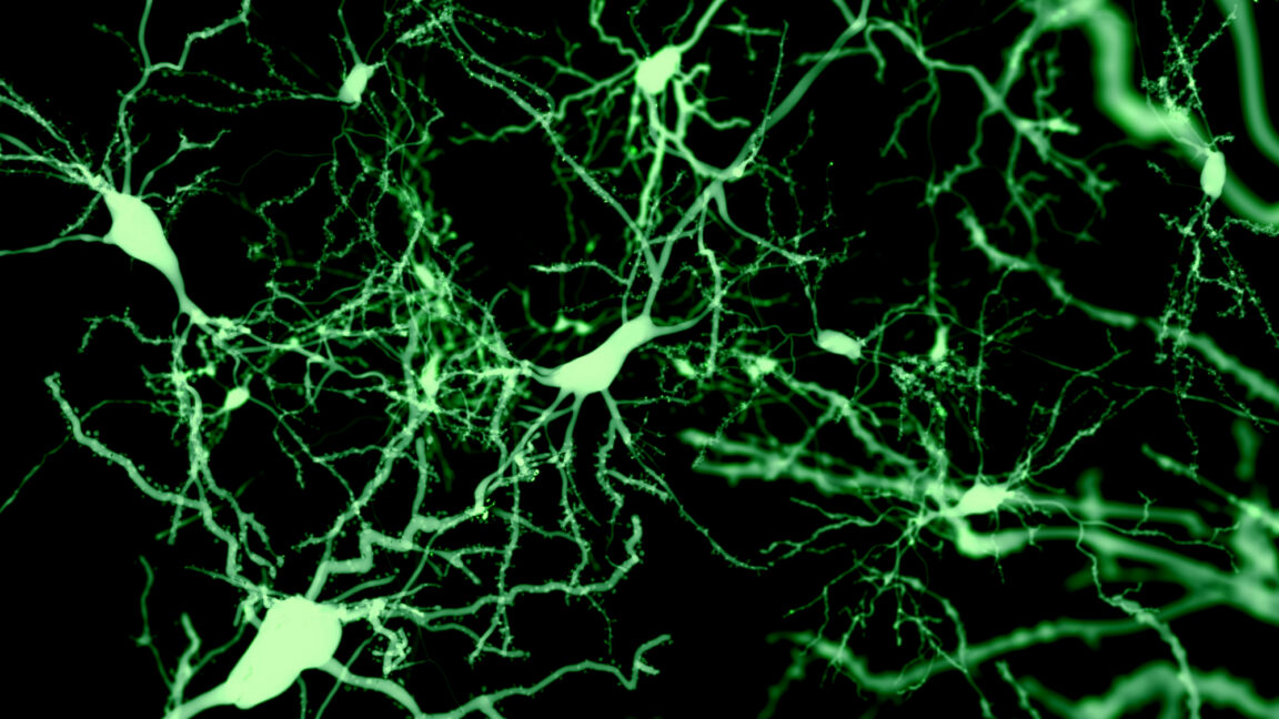
Sst-Chodl neurons sleep research has produced an unusually direct result in mice: artificially activating a rare set of inhibitory cells in the cerebral cortex increased sleep and shortened the time animals took to fall asleep. The work challenges the long-standing view that the cortex mainly receives sleep instructions from deeper brain structures, though it does not establish a treatment for people.
Geoffrey Terral and Renata Batista-Brito’s group at Albert Einstein College of Medicine reported the findings in a recent Nature study, according to Ars Technica. The experiments were conducted in mice, primarily in visual cortex.
What are Sst-Chodl neurons and how did they affect sleep?
Sst-Chodl neurons are inhibitory cortical neurons identified by activity of two genes, Sst and Chodl. Hitting either genetic marker alone would catch a broad and mixed set of cells, so the researchers developed a method that labels cells only when both genes are active, Ars Technica reported.
The cells are scarce. Inhibitory neurons account for about one-fifth of cortical neurons, while Sst-Chodl neurons represent roughly one in 1,000 cortical neurons, according to the report. Yet their wiring is extensive: rather than acting only near their cell bodies, they send long projections across and beyond the visual cortex.
Researchers imaged 111 of the cells while monitoring mice’s movement, pupil size, muscle tone and cortical electrical activity. Ninety-five were active during slow-wave sleep and quiet, motionless wakefulness, and were inactive during running and REM sleep, Ars Technica reported.
They then used optogenetics, a technique that gives selected cells a light-sensitive ion channel so scientists can trigger their firing with light. Stimulating Sst-Chodl neurons in visual cortex increased delta-wave power, sharpened the timing of neural spikes, and made cortical DOWN states, intervals of near-silence between bursts of activity, both more frequent and longer. Overall firing rates changed little. The apparent effect was coordination, not a blunt shutdown of the cortex.
In freely moving mice, stimulation across the cortex increased both slow-wave and REM sleep, reduced sleep-onset time, and prompted the animals to go to their nests during the day. It also produced sleep effects during the dark phase, when nocturnal mice are generally active.
How does this fit with established sleep biology?
The finding adds a cortical participant to a system already known to involve several regions. The National Institute of Neurological Disorders and Stroke says the hypothalamus contains neural control centers for sleep and wakefulness, while the brainstem helps govern transitions between the two. The hypothalamus’s suprachiasmatic nucleus also uses light information from the eyes to align circadian rhythms with the day-night cycle.
Batista-Brito and colleagues have proposed that Sst-Chodl neurons may sense homeostatic sleep pressure, the mounting drive to sleep after prolonged wakefulness. That remains a hypothesis. The researchers do not yet know what naturally activates the cells, and they were examining whether their connections differ in other cortical regions, including prefrontal cortex.
The clean reading of the evidence is narrower than a “sleep switch” headline: activating this rare cell population changed cortical rhythms and sleep behavior in mice. It does not show that the cells alone explain sleep, nor does it show a way to treat insomnia or alter human sleep safely.
This story draws on original reporting from Ars Technica.